Facial Care
Discover Our Aesthetic Treatments for the Face
ERYTHROSIS COLI - POIKILODERMA OF CIVATTE
The term "poikiloderma" refers to a skin condition characterized by atrophy with observed changes in hypo/hyperpigmentation and dilation of fine blood vessels (telangiectasia) on the affected skin.
This condition was first described in 1923 by a French dermatologist named Civatte.
ERYTHROSIS COLI - POIKILODERMA OF CIVATTE
Question 1: What are the clinical features of poikiloderma of Civatte?
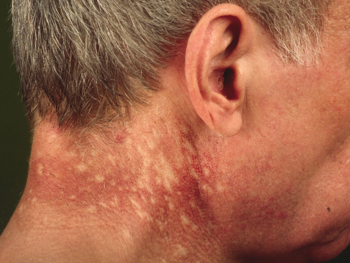
Poikiloderma of Civatte is characterized by the combination of redness and/or pigmentation (reddish-brown appearance) with atrophy (thin skin) that symmetrically affects sun-exposed areas such as the sides of the neck and cheeks. Poikiloderma of Civatte usually spares the shaded area under the chin.
Question 2: What are the complications of poikiloderma of Civatte?
Poikiloderma of Civatte is generally asymptomatic, but some patients may experience mild burning, itching, episodic vasomotor flushes, and sensitive skin in the affected area. No systemic involvement or serious complications are associated with poikiloderma of Civatte.
Question 3: Who is affected by poikiloderma of Civatte?
Poikiloderma of Civatte is more common in middle-aged and elderly individuals with fair skin, especially those who are heavily sun-exposed. The highest frequency is observed in menopausal women. The exact incidence is unknown as many patients have a mild form of the disease and may not necessarily seek medical attention.
Question 4: How is poikiloderma of Civatte diagnosed?
The diagnosis of poikiloderma of Civatte is made clinically. A punch biopsy may show typical histology with hyperkeratosis, epidermal atrophy, pigmentary incontinence, telangiectasias, variable superficial lymphohistiocytic infiltrate, and solar elastosis.
Question 5: What are the causes of poikiloderma of Civatte?
The exact cause is unknown. Long-term sun exposure is considered the main contributing factor. Other factors include: Fair skin (Fitzpatrick skin type I and II) Photosensitizing components in cosmetics and toiletries, especially fragrances Hormonal changes related to menopause or low estrogen levels Genetic predisposition.
WHAT IS THE TREATMENT FOR POIKILODERMA OF CIVATTE?
Despite adequate and well-conducted medical treatment, this condition remains difficult to completely erase. However, treatment with intense pulsed light (IPL) offers good results by greatly reducing the intensity of discolorations for a more comfortable aesthetic outcome.
The mixed erythematous and pigmented character of this elastotic skin makes IPL preferable to lasers in this indication. A highly intense light wave specifically targets the elements responsible for poikiloderma: hemoglobin (for diffuse redness) and melanin (for the brown appearance).
IPL is an effective, non-invasive, and minimally painful therapy for poikiloderma of Civatte. It provides reduction of pigmentation and telangiectasias with a low risk profile. Additional benefits include subjective changes in skin texture improvement.
Non-ablative fractional laser treatment can improve the vascular, pigmented, and textural components of poikiloderma of Civatte.
Preparations containing hydroquinone and alpha-hydroxy acids can help fade pigmentation.
The patient should be educated to avoid sun exposure and properly use broad-spectrum SPF 50+ sunscreen to prevent recurrences and avoid all perfumes on or near the affected area, including those contained in soaps.
HOW DOES A SESSION AT CLINIQUE BIOLASER TAKE PLACE?
- Each session lasts between 15 and 30 minutes.
- The treated area must be clean, dry, not tanned, and without makeup.
- Possible side effects may include increased redness and swelling for 4 to 6 hours, and brown crusts (do not scratch) that can last up to a week.
- There is no social downtime.
- It is necessary to apply a healing cream and sunscreen for a few days after each session.
- Generally, very satisfactory results are achieved after 2 to 6 sessions. We respect a 4 to 6-week interval between each session to allow the skin to rest and regenerate. The doctor may propose a two-step treatment if the skin has a combination of redness and brown spots (hyperpigmentation). This involves treating brown pigmentation first and then the dilated vessels responsible for diffuse redness.
- However, in some cases, a few additional sessions may be necessary for extensive or resistant lesions.
- An annual session will generally be required for maintenance.
